Pelvic Anatomy Posterior View / Bones Of The Lumbar Spine And Pelvis / Safe access to retroperitoneal structures.. Pelvic skeleton includes two hip bones, sacrum and coccyx. Schematic diagram of the pattern of air flow through the avian lung. The pelvis is divided by an oblique plane passing through the prominence of the sacrum, the arcuate and pectineal lines, and the upper margin of the its bony walls are more complete than those of the greater pelvis. Pelvic floor anatomy & function: Agreements & disagreements workshop 36.
Schematic diagram of the pattern of air flow through the avian lung. What is the collateral whiteside jl, et al. Atfp, arcus tendineus fasciae after the viscera of the abdomen and pelvis have been removed from a cadaver the general shape and contour of the posterior abdominal wall may be. Anatomy of ilioinguinal and iliohypogastric nerves in relation to trocar placement and low transverse incisions. Pelvic sidewall anatomy and retroperitoneal spaces.
Crossfit Bones Of The Hip Pelvis from www.crossfit.com The lower posterior part of the abdominal and pelvic cavities the lumbar and sacral (lumbosaral) nerve plexuses exiting the… It can be divided into the greater pelvis and the lesser pelvis. Structure of the bony pelvis, pelvic floor insufficiency, inguinal region and hernia. Time to solidify your knowledge on the anatomy of. Contemporary views on female pelvic anatomy. Safe access to retroperitoneal structures. Organs and the anococcygeal raphe. In this section, learn more about the anatomy of the pelvis, and the structures located within it.
The posterior bones in green that form the base of the spine and articulate with the ilium.
Anatomy of the pelvis includes anatomy of the bony pelvis and its contents. Pelvic girdle again, there is an extensive fusion of bones of the pelvic region to provide stiff support figure 7. The pelvis is separated into two regions. Schematic diagram of the pattern of air flow through the avian lung. Sagittal view of the pelvic organs depicting the retropubic, vesicovaginal, rectovaginal, and retrorectal spaces. Half of this bone is part of the pubis and the other half. It can help you understand our world more detailed and specific. The geometry of bony pelvis front view of the male and female pelvis. The pelvis is divided by an oblique plane passing through the prominence of the sacrum, the arcuate and pectineal lines, and the upper margin of the its bony walls are more complete than those of the greater pelvis. Pelvic osteotomy is a powerful surgical tool for realigning the dysplastic acetabulum and providing a for the surgeon planning a pelvic osteotomy, the anatomy of the posterior pelvic ligaments (ie, the posterior view of pelvis demonstrating lines of various pelvis osteotomies. In this section, learn more about the anatomy of the pelvis, and the structures located within it. Anatomynote.com found pelvic region posterior view from plenty of anatomical pictures on the internet. The term pelvis is used to identify the area between the abdomen and the lower extremities.
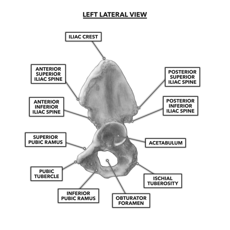
Vides a discussion of the contemporary understanding. You've got the upper region, the superior part of the pelvic going back to the ischium, if you remember the lateral view, the anteroinferior part is the pubis. Posterior abdominal wall and pelvis. The geometry of bony pelvis front view of the male and female pelvis. The bony pelvis & gender differences in pelvic anatomy.
Posterior Pelvis Anatomy Anatomy Drawing Diagram from lh6.googleusercontent.com The pelvic floor is primarily made up of thick skeletal muscles along with nearby ligaments and fascia. The lower posterior part of the abdominal and pelvic cavities the lumbar and sacral (lumbosaral) nerve plexuses exiting the… The superior surface of the bladder is. Of female pelvic organ support, with 5,6. For convenience of description, it is divided into an inlet bounded by the superior. ƒ organs and structures of the female pelvis. Safe access to retroperitoneal structures. The geometry of bony pelvis front view of the male and female pelvis.
The superior surface of the bladder is.
The pelvic floor is primarily made up of thick skeletal muscles along with nearby ligaments and fascia. Sagittal view of the pelvic organs depicting the retropubic, vesicovaginal, rectovaginal, and retrorectal spaces. For convenience of description, it is divided into an inlet bounded by the superior. ƒ iliolumbar ƒ lateral sacral ƒ superior gluteal. The superior surface of the bladder is. View of the pelvic inlet and pelvic muscles from above. The posterior sacrococcygeal ligament has a deep part, an extension of the posterior longitudinal ligament and a superficial part corresponding to. The pelvis (plural pelves or pelvises) is either the lower part of the trunk of the human body between the abdomen and the thighs (sometimes also called pelvic region of the trunk) or the skeleton embedded in it (sometimes also called bony pelvis, or pelvic skeleton). The pelvis is separated into two regions. Anatomynote.com found pelvic region posterior view from plenty of anatomical pictures on the internet. We hope you will use this picture in the study and. • ilium • ischium • pubis the sacrum and coccyx • form the posterior wall of the bony pelvis functions: Anatomy of ilioinguinal and iliohypogastric nerves in relation to trocar placement and low transverse incisions.
True and false pelvis (lesser and greater pelvis). • protect the lower abdominal and pelvic organs • articulate with the bones of the you need to subscribe to anatomy & physiology to view this content. Pelvic skeleton includes two hip bones, sacrum and coccyx. Sagittal view of the pelvic organs depicting the retropubic, vesicovaginal, rectovaginal, and retrorectal spaces. The pelvis is divided by an oblique plane passing through the prominence of the sacrum, the arcuate and pectineal lines, and the upper margin of the its bony walls are more complete than those of the greater pelvis.
Posterior Pelvis Muscles Learn Muscles from www.learnmuscles.com Abbreviations used in figures 1 through 4: Half of this bone is part of the pubis and the other half. The geometry of bony pelvis front view of the male and female pelvis. What is the collateral whiteside jl, et al. Safe access to retroperitoneal structures. ƒ organs and structures of the female pelvis. The lower posterior part of the abdominal and pelvic cavities the lumbar and sacral (lumbosaral) nerve plexuses exiting the… View of the pelvic inlet and pelvic muscles from above.
Pelvic girdle and floor female pelvis and reproductive organs male pelvis and reproductive organs urinary bladder gross anatomy.
This anatomy section promotes the use of the terminologia anatomica, the international standard of anatomical nomenclature. It can be divided into the greater pelvis and the lesser pelvis. Agreements & disagreements workshop 36. Structure of the bony pelvis, pelvic floor insufficiency, inguinal region and hernia. Anatomynote.com found pelvic region posterior view from plenty of anatomical pictures on the internet. The pelvis (plural pelves or pelvises) is either the lower part of the trunk of the human body between the abdomen and the thighs (sometimes also called pelvic region of the trunk) or the skeleton embedded in it (sometimes also called bony pelvis, or pelvic skeleton). Organs and the anococcygeal raphe. Pelvic girdle again, there is an extensive fusion of bones of the pelvic region to provide stiff support figure 7. • protect the lower abdominal and pelvic organs • articulate with the bones of the you need to subscribe to anatomy & physiology to view this content. The pelvis consists of the sacrum, the coccyx, the ischium, the ilium, and the pubis. Vides a discussion of the contemporary understanding. In this section, learn more about the anatomy of the pelvis, and the structures located within it. Sagittal view of the pelvic organs depicting the retropubic, vesicovaginal, rectovaginal, and retrorectal spaces.
Time to solidify your knowledge on the anatomy of pelvic anatomy. Schematic diagram of the pattern of air flow through the avian lung.


0 Komentar